What is MRI of the Breast?
.jpg) Magnetic resonance imaging (MRI) is a noninvasive medical test that physicians use to diagnose and treat medical conditions.
Magnetic resonance imaging (MRI) is a noninvasive medical test that physicians use to diagnose and treat medical conditions.
MRI uses a powerful magnetic field, radio frequency pulses and a computer to produce detailed pictures of organs, soft tissues, bone and virtually all other internal body structures. MRI does not use ionizing radiation (x-rays).
MRI of the breast offers valuable information about many breast conditions that cannot be obtained by other imaging modalities, such as mammography or ultrasound.
How should I prepare?
Someone from the MRI department usually attempts to contact you a day or two before your appointment to go through a safety screening due to the strong magnetic field associated with this exam. You will be asked about any previous surgeries and/or implants that you have. Although most implants are safe for the MRI environment, there are some implants that need to be researched before the exam to make sure the correct parameters are used to avoid any issues. Some implants are unsafe and you will not be able to have an MRI. Jewelry and other accessories should be left at home if possible, or removed prior to the MRI scan because they can interfere with the magnetic field of the MRI unit, metal and electronic items are not allowed in the exam room.
Breast MRI examinations also require you to receive an injection of contrast material into the bloodstream. The technologist or a nurse will ask if you have allergies of any kind, such as an allergy to contrast material, drugs, food, etc. They will also ask questions that pertain to kidney health. Some conditions, such as severe kidney disease, may prevent you from being able to receive gadolinium therefore you cannot have a breast MRI. It is a good idea to hydrate before your MRI as this will help with venous access and also very important to hydrate after the MRI to help flush the contrast out of your body.
If you have claustrophobia (fear of enclosed spaces) or anxiety, you may want to ask your physician for a prescription for a mild sedative prior to your scheduled examination.
How is the exam performed?
In general, you will be in the MRI department approximately 45 minutes. The time that is spent on the exam table performing the MRI scan is about 25 minutes. The 25 minutes is separated into several sets each lasting various times. The technologist will talk with you between each of the sets. It is very important during a breast MRI to remain very still and to not move out of position until the scan is complete. It is extremely important for interpretation that all of the breast anatomy is in the same spot for each of the sequences. If there is motion on the images or the position has changed during the exam it may render the MRI uninterpretable.
 You will be asked to wear a gown during the exam. No clothing with metal can be worn during a breast MRI. Guidelines about eating and drinking before an MRI exam vary with the specific exam and also with the imaging facility. Unless you are told otherwise, you may follow your regular daily routine and take food and medications as usual.
A nurse or technologist will start an IV so that the contrast can be injected with a power injector at a specific rate and timing half way through the exam.
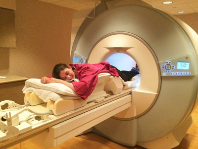
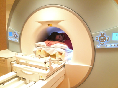
You will be asked to lie on your stomach for the exam with your breasts in a cupped device. There is no compression of the breasts during a routine MRI as with a mammogram. You will be required to wear headphones or earplugs during the MRI due to the noise that is created by the MRI machine. It is normal for the area of your body being imaged to feel slightly warm, but if it bothers you, notify the technologist. When the contrast runs into your vein and throughout your body you may experience sensations such as coolness or trickling in your arm, followed by warmth or flushing in your face, chest, abdomen and pelvis. Some people even experience a very brief moment of nausea or a strange taste or smell. All of these feelings are actually normal and pass within a minute or two. However if you cannot continue with the test you will have a way to signal the technologist to let them know to stop the exam (either visually or with an alarm button).
 The bore size for the MRI machine at Wilton Medical Arts is 70 cm in diameter (27.5 in) and approximately 4 feet long. Patients are put into the machine feet first and positioned so that their chest is in the center of the bore. Your head does go inside the machine but is very close to the outside opening.
Patients are generally positioned face down and arms up as seen in the upper right picture which allows for headphones to listen to music. However, if patients find this uncomfortable, the head can be turned to the side on pillows with earplugs for hearing protection instead of headphones.
Arms are usually raised above the head but can also be positioned as seen in the upper left picture if more comfortable.
If you have additional questions, please call Wilton Medical Arts MRI Dept. at 518-580-2262.
