
The Saratoga Hospital Center for Breast Care providers coordinate care to complete the diagnostic evaluation and provide treatment for breast disease.
The Women’s Imaging Center at Wilton Medical Arts has been designated a Breast Imaging Center of Excellence by the American College of Radiology.
![]() This prestigious designation is only awarded to centers that offer comprehensive services and meet strict quality standards for all breast breast imaging modalities (mammography, breast ultrasound, breast MRI and stereotactic breast biopsy).
This prestigious designation is only awarded to centers that offer comprehensive services and meet strict quality standards for all breast breast imaging modalities (mammography, breast ultrasound, breast MRI and stereotactic breast biopsy).
Breast cancer screening is done to detect breast cancer before there are any symptoms. Finding breast cancer early gives the best options for treatment and cure.
When an abnormality is detected on a screening test, additional diagnostic tests and a biopsy may be required.We also perform screening mammograms at Saratoga Medical Park in Malta. Conveniently located off of the Northway at Exit 12, the Malta Med Emergent Care building offers medical imaging and additional specialty services on the second floor.
Why Getting a Screening Mammogram Matters
BREAST IMAGING
Mammography
Mammography uses x-rays to produce detailed images of the breast, which are viewed on a high-resolution computer monitor. A board-certified radiologist will analyze the images, describe any abnormalities and make a diagnosis. A report is sent to the referring physician and the patient is also notified of the results.
When to Start Having Mammograms
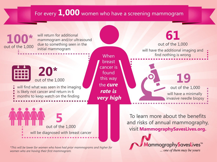
As you can see in the graphic above, for every 1,000 women who have screening mammograms, about 100 women will be called back for additional testing and 19 will have a breast biopsy, but fortunately only 5 will be found to have breast cancer. The most significant risk associated with screening mammography, therefore, is a “false positive” result that can lead to further evaluation but does not ultimately identify a cancer.
In part because breast cancer is more common in older women, and because there is concern about false positive screening tests, some of the guidelines for screening mammography have recently become more nuanced. The United States Preventative Services Task Force (USPSTF) now recommends that women with average risk start regular mammography at age 50. Their guidelines indicate that the decision to start screening mammography at age 40 should be an individual one, balancing the potential harms of false positive results against the benefits of early cancer detection.
The American Cancer Society recommends screening mammography start at age 45, but also recommends that women be offered the choice to start screening mammography at age 40. That American College of Obstetricians and Gynecologists, the American College of Radiology, and the National Comprehensive Cancer Network still all recommend that screening mammography start at age 40.
The guidelines which offer women the option to start screening mammography later are all for average risk women. Women with increased risk, due to a family history of breast cancer or other risk factors, are more likely to benefit from early screening and should definitely start mammography at age 40.
Digital Breast Tomosynthesis (3D mammography)
Digital Breast Tomosynthesis generates multiple images through the breast tissue and can more effectively screen for invasive tumors, especially for women with dense breast tissue.
Breast Ultrasound
Breast Ultrasound can be used to further evaluate dense breast tissue seen on mammogram or abnormalities felt on breast examination. Ultrasound uses sound waves to produce images, and can help the radiologist determine if a breast mass is solid or a fluid-filled cyst.
Magnetic Resonance Imaging (Breast MRI)
Magnetic Resonance Imaging cas an aid to mammography and ultrasound imaging. It can be used to evaluate the extent of known disease, measure the effectiveness of chemotherapy, and assess for implant rupture. Breast MRI is also appropriate for screening patients who are at a high risk of developing breast cancer. Intravenous contrast is administered for breast MRI exams, except when evaluating implants. Breast MRI has the advantages of not being limited by tissue density, being extremely sensitive, and not using x-rays. It can, however, highlight areas of benign fibrocystic tissue and give false positive results.
Read more here »
BREAST BIOPSIES
Needle Biopsies
An image guided needle biopsy is a highly accurate alternative to a surgical breast biopsy. This procedure is done by the radiologist, using local anesthesia to numb the area while small pieces of tissue are obtained through a needle. The image guidance is done using the modality by which the radiologist can best see the abnormality:
- A stereotactic core needle biopsy is done using x-ray guidance
- An ultrasound guided needle biopsy is done using ultrasound to guide the needle
- MRI guided needle biopsy is done with MRI
The tissue samples are sent to pathology for analysis, and the patient is called with the biopsy results as soon as they are available (typically 3 – 5 days).
Surgical (Excisional) Biopsies
Although it is our preference to offer patients minimally invasive image guided needle biopsies, there are occasions when that approach is not technically feasible or appropriate. A surgical biopsy, also known as an excisional biopsy, can be done by your surgeon, in the operating room.
Wilton Medical Arts Building
3040 Rte 50,
Saratoga Springs, NY 12866
Phone: 518-580-2170
Fax: 518-580-2171
Hours
Monday - Friday
8:00 a.m. to 4:30 p.m.
Women's Imaging Contact Info »